Thursday, 2 October 2014

foods for ur brain


Can your diet make you smarter?


You bet. Research shows that what you eat is one of the most powerful influences on everyday brain skills, says Cynthia Green, PhD, founder and director of the Memory Enhancement Program at Mount Sinai School of Medicine in New York City and author of Brainpower Game Plan. Plus, the right foods may stave off Alzheimer’s and other forms of dementia. The key? Rounding out your meals with key nutrients that (along with exercise and daily brain games like these) keep brain cells healthy and prevent brain-damaging inflammation. “Your memory, attention span, and ability to learn will benefit from the healthful foods you choose,” says Green.

Fatty Fish
Seafood like salmon, albacore tuna, mackerel, and sardines are packed with omega-3 fatty acids, powerful and versatile nutrients that are essential for a healthy mind. About 40% of the fatty acids in brain cell membranes are DHA, one of the main omega-3 fatty acids in fish oil. Experts believe it’s probably necessary for transmitting signals between brain cells.
Researchers at Tufts University found that people who ate fish 3 times a week and had the highest levels of DHA in their blood slashed their risk of Alzheimer’s disease by 39%.

Eat it: At least twice a week (limit albacore tuna to no more than 6 ounces a week to minimize mercury exposure). Try these 7 delicious fish recipes.
Leafy Green and Cruciferous Veggies
Pile salads, stir-fries, and side dishes with broccoli, cauliflower, cabbage, kale, bok choy, and brussels sprouts. They’re filled with antioxidants like vitamin C and plant compounds called carotenoids, which are particularly powerful brain protectors.

Antioxidants prevent damage from free radicals, which are waste products your body makes when cells use fuel to create energy. Your brain is especially vulnerable to damage from free radicals because it uses a lot of fuel (it’s only about 3% of your body weight but uses up to 17% of your energy). Since your mind makes a lot of these toxic by-products, ample antioxidants help to disarm and defuse them.

While all antioxidants (from a variety of plants) are good for your brain, these cruciferous veggies are especially effective. A Harvard Medical School study of more than 13,000 women found that those who ate the most lowered their brain age by 1 to 2 years.

Eat it: Daily, as part of a well-rounded mix of other colorful veggies.
Avocado, Oils, Nuts, and Seeds
They all contain another important antioxidant: vitamin E. In one study, researchers found that people who consumed moderate amounts vitamin E—from food, not supplements—lowered their risk of AD by 67%.

Eat it: Frequently; shoot for 15 mg of E a day, the equivalent of 2 ounces of almonds.

Chocolate
Sweeten your brain-boosting diet with the dark kind (at least 70% cocoa); it contains flavonoids, another class of antioxidants that some research links to brain health. Other flavonoid-rich foods include apples, red and purple grapes, red wine, onions, tea, and beer.
Eat it: Frequently, as part of a healthy total calorie intake. Up to half an ounce daily has also been shown to lower blood pressure.

Curry
Go for Thai or Indian takeout; these cuisines often use the potent spice known to fight inflammation. Animal studies have shown that curry’s active ingredient, curcumin, actually clears away Alzheimer’s-causing proteins in the brain called amyloid plaques.

Eat it: As an ingredient in pasta sauces, salad dressings, or meat marinades. (Consider these tasty recipes that make Indian food easy to make.)

Berries
Research indicates these antioxidant powerhouses may protect your brain, although the mechanism isn’t fully understood. Some scientists think they help to build healthy connections between brain cells.

Eat them: Daily, added to yogurt, oatmeal, or cereal for breakfast or an afternoon snack.
Whole grains

Monday, 29 September 2014

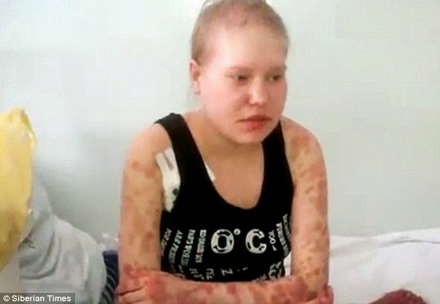
krokodil A flesh eating drug


In the vast pharmacopeia of illegal street drugs, few have as heinous a reputation as desomorphine, popularly known as crocodile or "krokodil." An opiate that's chemically related to morphine and heroin, krokodil earned its name in two ways: Addicts develop dark, scaly lesions on their skin, and the drug tends to eat its victims alive, like a crocodile.


Krokodil first surfaced in Russia several years ago, where users discovered the drug was much cheaper than heroin and could easily be cooked in a kitchen by combining codeine with gasoline, oil, alcohol or paint thinner, Fox News reports.

And now, krokodil has arrived in the United States: Two cases of people addicted to the drug have been reported in Arizona. "As far as I know, these are the first cases in the United States," said Dr. Frank LoVecchio, director at Banner Good Samaritan Poison and Drug Information Center in Arizona, as quoted by Fox News. "So we're extremely frightened." [Trippy Tales: The History of 8 Hallucinogens]


Krokodil is injected with a hypodermic needle, and necrosis — the death and decay of living tissue — can quickly set in. Gangrene and amputations are common among addicts, who often die within two or three years after they start using the drug, according to the Daily Mail.
Like other opiates, krokodil is powerfully addictive, and even those users who kick the habit often walk away severely disfigured: severe scarring, bone damage, amputated limbs, speech impediments, poor motor skills and varying degrees of brain damage — for life.


And it doesn't take much lab equipment to make: The illegal production of krokodil in Russia and the Ukraine is similar to the chemical processes used to home-produce methamphetamine, according to an article published online June 3 in the International Journal of Drug Policy. To make krokodil, producers transform codeine into its opiate analog called desomorphine, whose analgesic effect is about 10 times greater than that of morphine — it's also about three times more toxic than morphine, the authors wrote.

Krokodil users may avoid seeking help partly due to the stigma attached to drug use. "In countries where public campaigns or media position drug use as social evil and where health providers are viewed as closely aligned with law enforcement or other systems of social control (e.g. child protection agencies), PWID [people who inject drugs] are likely to postpone seeking treatment for medical problems that need urgent professional care," the authors wrote in their journal article.

"This is really frightening," Dr. Aaron Skolnik, a toxicologist at Banner Good Samaritan Poison and Drug Information Center in Phoenix, told Fox News. "This is something we hoped would never make it to the U.S. because it's so detrimental to the people who use it."

Saturday, 27 September 2014

secrets of longevity


Blood Of Oldest Woman In The World Reveals Secrets Of Longevity, Suggests We Can Live Longer


A study on the blood of the world's oldest woman has revealed that the secret to longevity is in our white blood cells. Photo courtesy of Shutterstock
It seems that 500 years after the death of Juan Ponce de Leon, science may have just discovered the mysterious fountain of youth. Only it’s not a fountain, and it’s definitely not in Florida. He actually had it all along — we all do. The secret to our longevity lies in our white blood cells. A study of blood and tissue samples from Hendrikje van Andel-Schipper, once the oldest woman in the world, revealed that death from old age is specifically caused by exhaustion of blood stem cells. Now that scientists have discovered what causes death, it automatically raises the question of what to do with this information. Some believe that manually replenishing these stem cells will result in the ability to fool Mother Nature and perhaps make death no longer inevitable.

Secrets in Blood

Born in 1890, van Andel-Schipper was known for her remarkable health in old age. She had a clear mind and disease-free body until her death in 2005. As she wished, upon her death van Andel-Schipper’s body was donated to science, and her blood and tissue samples helped scientists understand how the body is affected by old age. What scientists found is that our lifespan is limited by the capacity for stem cells to replenish, New Scientist reported. When stem cells are eventually no longer able to self-replenish, they gradually die off. The individual will no longer be able to replace tissue, and cells will soon succumb to a natural death of old age.

At the end of her life, two thirds of van Andel-Schipper’s remaining white blood cells had originated from just two stem cells. This suggests that most or all of the blood stem cells she had started with in life had burned out and died. Also, the telomeres on van Andel-Schipper’s white blood cells were drastically worn down. Telomeres are the protective tips on chromosomes that burn down like a candle wick each time a cell divides. During life, the number of active stem cells shrinks, and their telomeres shorten to the point at which they die. This point is called stem-cell exhaustion.

'Fountain of Youth'?

Does understanding the cause of death mean that we will be able to one day control death? Some scientists believe it may be possible to rejuvenate aging bodies with re-injections of stem cells saved from birth or early life. “If I took a sample now and gave it back to myself when I’m older, I would have long telomeres again—although it might only be possible with blood, not other tissue,” Henne Holstege, lead researcher of the study on van Andel-Schipper’s blood, told New Scientist. Rather than using this information to create a theoretical "fountain of youth," scientists hope that the results can be used in correlation with studies on Alzheimer’s disease to reveal why some people are more susceptible to the disease at an early age.

Van Andel-Schipper’s blood also gave some important insight into our bodies’ natural defenses against cancer. Researchers found that although there were many mutations in van Andel-Schipper’s blood, they were all harmless. These mutations were the result of mistaken replication of DNA, but due to their absence of cancer, researchers concluded that van Andel-Schipper had an advanced system for repairing or aborting cells with dangerous mutations. “When there is a mutation, there’s an opportunity for selection and some somatic mutations lead to cancer. Now we see the range of somatic mutations in normal, non-cancerous tissues like blood, so we can start to think about health consequences,” said Chris Tyler-Smith of the Wellcome Trust Sanger Institute in Hixton, Texas.

Friday, 26 September 2014

1.4 Million People Might Contract Ebola By February why think for a while


Why The CDC Thinks 1.4 Million People Might Contract Ebola By February
A crash course in modeling the outbreak of a deadly disease


Last June, the Kenema Government Hospital in Sierra Leone built a temporary ward in anticipation of an influx of Ebola patients. The makeshift building was covered with thin, tin metal sheets, haphazardly held to the walls by wire twist-ties. Robert Garry says that when the structure fell, it seemed inevitable.


But finding physical space for patients wasn’t the only problem at the hospital. Even after the ward was rebuilt, Garry and his staff have been overworked, and many have contracted the disease. Another doctor at the Kenema Hospital, Sheik Humarr Khan, was often left as the only person caring for 80 people. But he told his sister: “If I refuse to treat them, who would treat me?”

Western Africa is struggling to prevent collapse during the worst Ebola outbreak ever. This week the CDC released a study predicting that by January of 2015, 1.4 million people may have contracted the virus. With new studies suggesting that the virus's mortality rate may be close to 70 percent, in the next six months 980,000 more people might die from the disease. Thomas Gift, one of the people who worked on the CDC report, explained that their model was based on daily case counts from the current outbreak, as well as estimates of previous ones.

To develop these types of predictive models, epidemiologists break individuals up into four groups: people who are at risk, people who are infected but not contagious, people who are infected and contagious, and people who have recovered. Ultimately, researchers try to determine how those groups interact with each other and predict movements between the groups. These movements comprise what is known as the “effective reproductive rate,” or the number of people each infected person will give the disease to. This number is called R. When R is less than 1, the outbreak is declining; if it’s more than 1, the disease is spreading.


Reports vs. Predicitions Graphs show how model predictions and actual case reports match up.
Gift says the parameters in the CDC study were set to minimize the differences between the model and what is being reported on the ground in Sierra Leone and Liberia. But he says, “Like all models, they are simplifications of reality.” Any model is only as good as the data that is fed into it, and it's possible that as many as 75 percent of all Ebola cases are going unreported.

For this reason, the model uses a correction factor to account for underreporting. The CDC study decided that factor should be based on the number of beds in use, since counting physical objects is somewhat more reliable than depending on case reporting. So researchers compared the number of patients in beds predicted by the model on August 28th to the number of actual patients in beds. But the authors only had access to the bed counts in Liberia, so it’s still just an estimate.

CDC scientists aren't the only ones trying to develop models of Ebola transmission. Christian Althaus, an epidemiologist at the University of Bern, has also developed an estimate of the outbreak. He says that the CDC’s estimate of 1.4 million is based on an assumption that the real number of cases is 2.5 times higher than reported. However, Althaus says that way before Ebola patients number in the millions, "we will see a dramatic change in behavior and probably a collapse of the countries. That might limit the spread to some extent, but it will be very important to maintain social order.”

He says it’s hard to quantify the impact of international aid, but Caitlin Rivers, a PhD student of computational epidemiology at Virigina Tech,  says her modeling results suggest that contact tracing and improving infection control can help but not halt the progress of the epidemic. “But there's no question it needs to be a top priority for the global public health community,” Rivers says.

Despite the uncertainty involved in developing these models -- including the chronic underreporting, which has made solid data about the outbreak difficult to come by -- even conservative estimates put the number of people infected by Ebola in the tens of thousands by the end of the year.

As of September 24th, WHO has reported 6,263 cases of infection, with 2,917 deaths. “The number of lives already lost is staggering, and grows every day,” Rivers says.

Tuesday, 23 September 2014

Aminocentesis tests problems


Amniocentesis

Your family / personal medical history or perhaps an ultrasound result may suggest to your doctor that you have a slightly greater chance of giving birth to an infant with a birth defect. Therefore your doctor may suggest that you undergo an amniocentesis, a test used to detect some foetal abnormalities.

What Is An Amniocentesis?

An amniocentesis is a test in which a small sample of liquor (water) is taken from around the developing baby. A needle is passed through the abdomen, the wall of the uterus (womb) and into the water (liquor) surrounding the baby without touching the baby or the placenta. Approximately 15mls of fluid is taken, this amount being small compared with the total amount of fluid present (at least 150mls) at the time of the test at 16 weeks.

Who Is Offered One?

Women of 37 years and over at the projected time of birth
Women who have already had a child with a problem such as Down’s Syndrome or Spina Bifida
Occasionally amniocentesis is done for other reasons. These include parents who are known to have a chromosomal abnormality or who are known to be at risk of having a baby with one of a number of rare abnormalities of their metabolism.

Why Are Women Over The Age Of 37 Offered An Amniocentesis?

As the age of the mother increases the chances of her having a baby with a chromosomal abnormality also increases. The most common chromosome abnormality is Downs Syndrome. Between 37 and 40 years of age the chance of having a baby with an abnormality is approximately 1 in 100. Beyond the age of 40 years this chance increases even further. The incidence of Spina Bifida does not seem to be correlated with age.

When Is Amniocentesis Performed?

An amniocentesis is usually performed from 14 weeks gestation.
What Are The Potential Complications?
There is a risk of miscarriage following this test, which is quoted at approximately 1 in 200 tests performed.

What Will Happen On The Day?

It is important that the mother’s bladder is full; this lifts the uterus up out of the pelvis making it easier to see the baby with ultrasound. The ultrasound scan is performed prior to the amniocentesis so that the pregnancy and the placenta can be checked and to determine whether there is a singleton or twin pregnancy. The ultrasound scan is quite painless and merely involves spreading a little warm gel over the lower abdomen and then moving an ultrasound probe gently over the skin surface.
After the ultrasound scan has been performed, the abdomen is cleaned and the needle is inserted into the uterus. The needle is watched all the time on the ultrasound screen. It then only takes about one minute to draw up 15-20mls of fluid into a syringe. After removing the needle and again checking the baby, the mother is able to get dressed and go home.
Almost without exception, patients experience far less discomfort from the test than they had expected. Any discomfort felt is usually similar to that felt for a simple blood test. It is recommended that for the rest of the day things are taken quietly, but after this to return to a usual routine.


Could There Be Any Problems Afterwards?

It is very rare to have any problems at all after the test. Occasionally there may be some mild discomfort due to a little bruising under the skin. If there is any loss of blood or water from the vagina or any pain after the amniocentesis you should consult your obstetrician.

What Tests Are Performed On The Fluid Taken?

An estimate of the amount of a particular protein (alpha feto-protein). There are usually large amounts of this protein present in the fluid if a baby has a defect called Spina Bifida.
The second test is carried out on the cells which are present in the fluid. The chromosomes of these can be examined and in this way it is possible to detect whether the baby has Downs Syndrome or any other less common chromosome problems. The sex of the baby is also determined.

Saturday, 20 September 2014

weekly injectable insulin by FDA


FDA approves weekly injectable diabetes drug